
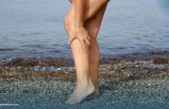
Venous reflux, also known as chronic venous insufficiency, occurs when one-way valves in the leg veins weaken and fail to close properly, allowing blood to flow backward and pool in the lower limbs. This raised venous pressure often causes leg heaviness, swelling, cramping, and visible varicose veins.
Left untreated, venous reflux may progress to skin discolouration or venous ulcers. Early diagnosis with duplex ultrasound, combined with minimally invasive treatments such as endovenous radiofrequency ablation, can restore healthy circulation and help prevent long-term complications.
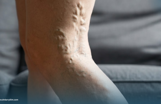
Varicose veins are enlarged, twisted veins that appear bluish or rope-like beneath the skin, most often affecting the legs. They develop when vein valves weaken and allow blood to pool, leading to heaviness, aching, swelling, and visible bulging veins that can affect comfort and confidence.
While often viewed as a cosmetic concern, varicose veins are a genuine vascular condition that can worsen over time. Treatment options range from compression therapy to minimally invasive procedures like sclerotherapy or endovenous ablation, restoring both vein health and appearance.
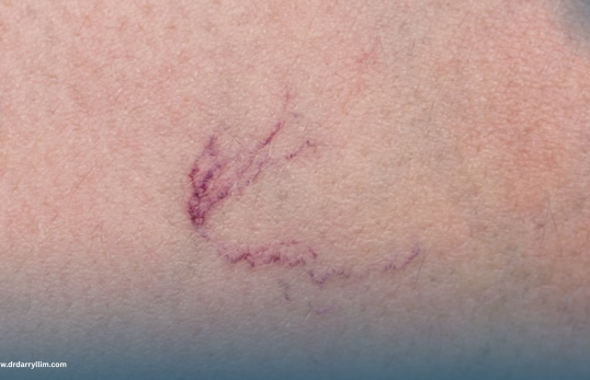
Spider veins are small, dilated blood vessels that appear as thin red, blue, or purple lines just beneath the skin’s surface, commonly on the legs and face. They form when tiny vein valves weaken, causing blood to pool in superficial vessels visible through the skin.
Though usually harmless, spider veins can cause mild aching or itching and are often a cosmetic concern. Treatments such as sclerotherapy or laser therapy can fade their appearance safely, and addressing underlying venous insufficiency helps prevent new ones from forming.
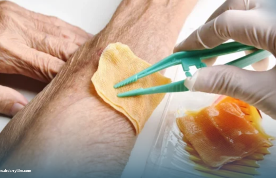
Poorly healing wounds are cuts, ulcers, or sores that fail to close within the expected timeframe, often due to underlying issues such as poor circulation, diabetes, or venous insufficiency. These wounds can become painful, prone to infection, and affect daily life.
Effective management addresses both the wound itself and its root cause, combining specialised dressings, infection control, and treatments to improve blood flow. Our advanced wound care approach aims to promote faster, more complete healing and reduce recurrence.
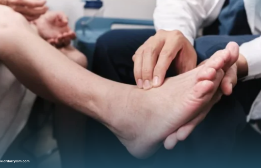
Peripheral vascular disease (PVD) occurs when narrowed or blocked arteries reduce blood flow to the limbs, most commonly the legs. This can cause pain while walking, numbness, cold extremities, and slow-healing wounds, and may signal wider cardiovascular risk.
Diagnosis typically involves imaging and blood flow studies, while treatment ranges from lifestyle changes and medication to minimally invasive procedures like angioplasty. Early management helps preserve mobility and reduce the risk of serious complications.
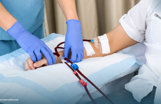
Dialysis access refers to the surgically created vein or graft, such as an arteriovenous fistula, that allows patients with kidney failure to safely undergo haemodialysis. Reliable access is essential for effective, long-term dialysis treatment and quality of life.
Complications such as narrowing, clotting, or infection can affect access function over time. Regular monitoring and minimally invasive procedures help maintain healthy access, ensuring dialysis continues smoothly with minimal disruption to care.
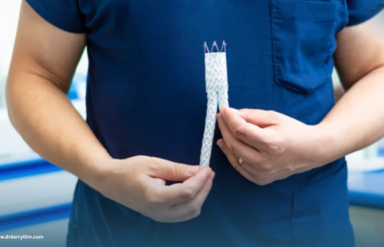
An aortic aneurysm is an abnormal bulge in the wall of the aorta, the body’s largest artery, which can grow silently over time without symptoms. If left untreated, an aneurysm risks rupture, a life-threatening emergency requiring immediate care.
Regular screening through ultrasound or CT imaging allows early detection, especially in at-risk individuals. Depending on size and growth rate, treatment may involve monitoring or minimally invasive endovascular repair to secure the aorta and prevent rupture.

Pelvic congestion syndrome occurs when veins in the pelvis become enlarged and engorged, similar to varicose veins elsewhere in the body. It is a common but often under-recognised cause of chronic pelvic pain, particularly among women.
Symptoms typically include a dull, aching pelvic pain that worsens with prolonged standing or before menstruation. Diagnosis involves imaging studies, and treatment often includes minimally invasive embolisation to close the affected veins and relieve symptoms.
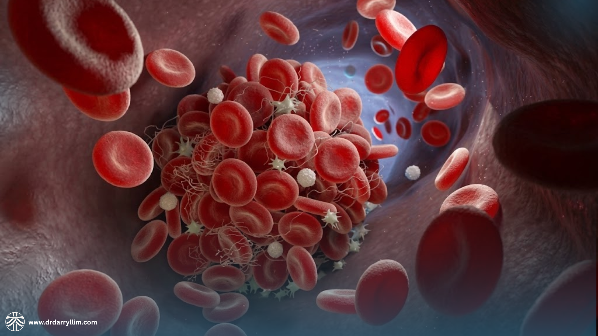
Deep vein thrombosis (DVT) occurs when a blood clot forms in a deep vein, most often in the leg, potentially causing swelling, pain, warmth, and redness. If part of the clot breaks free, it can travel to the lungs, causing a serious pulmonary embolism.
Risk factors include prolonged immobility, surgery, and certain medical conditions. Diagnosis relies on ultrasound imaging, while treatment typically involves blood-thinning medication and, in select cases, minimally invasive clot removal procedures.
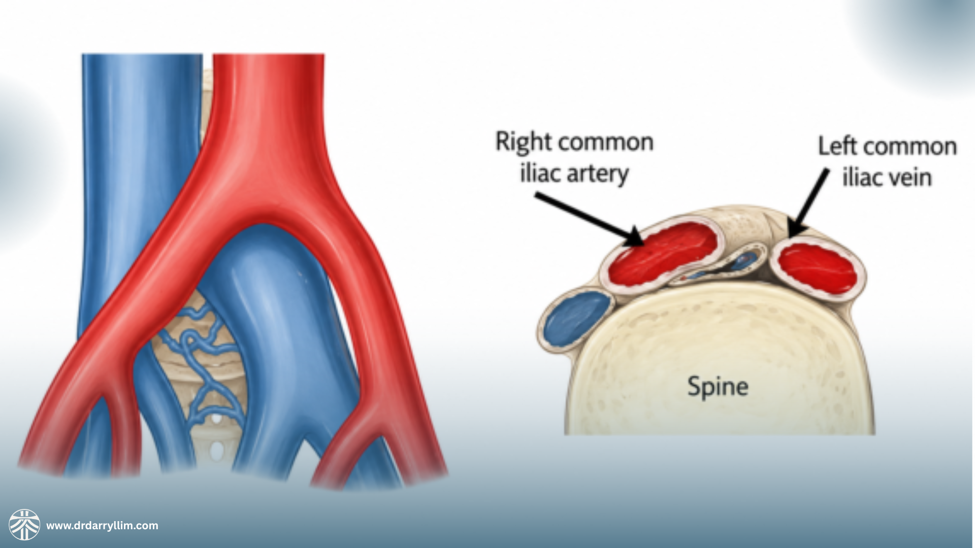
May Thurner syndrome occurs when the right iliac artery compresses the left iliac vein against the spine, narrowing it and restricting normal blood flow from the leg. This increases the risk of left-sided deep vein thrombosis and chronic leg swelling.
The condition is often under-diagnosed until a clot occurs. Imaging such as venography can confirm the compression, and treatment may involve blood thinners together with minimally invasive stenting to relieve pressure and restore healthy flow.